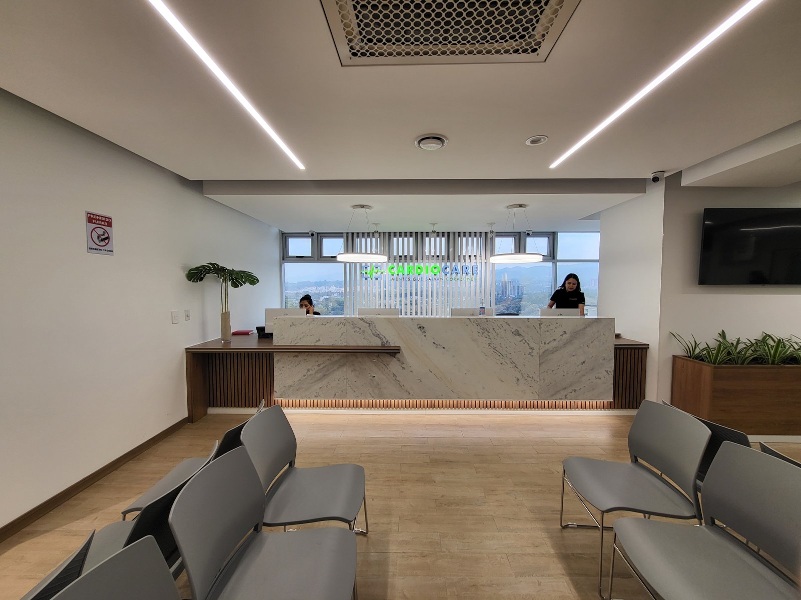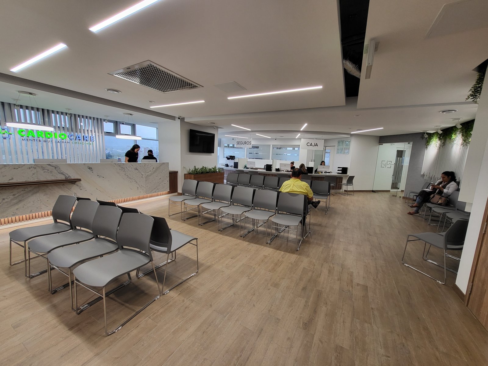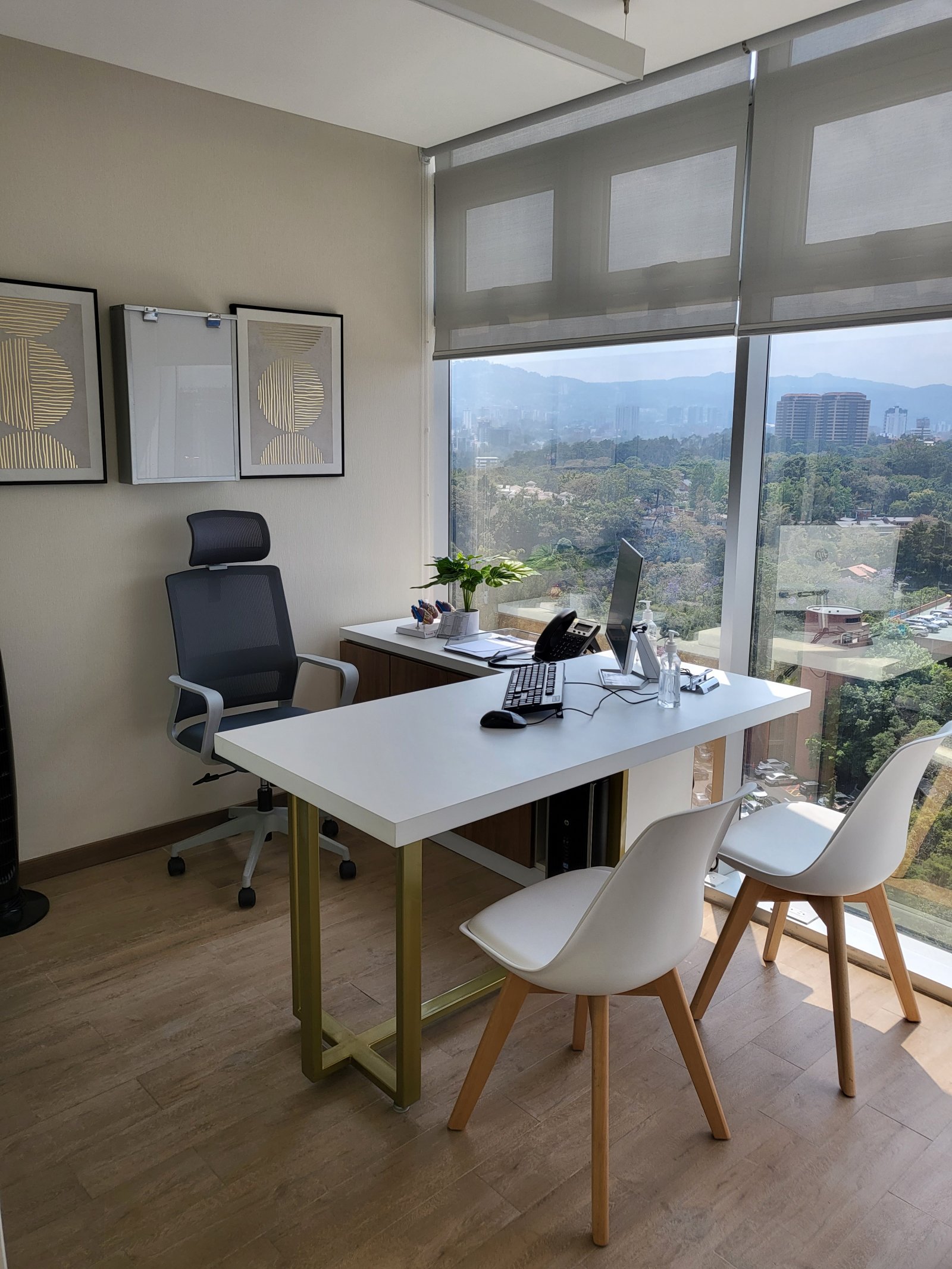


Expertos en corazón
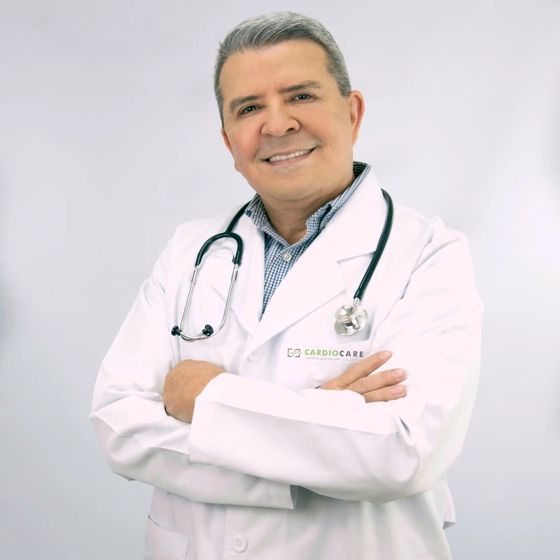
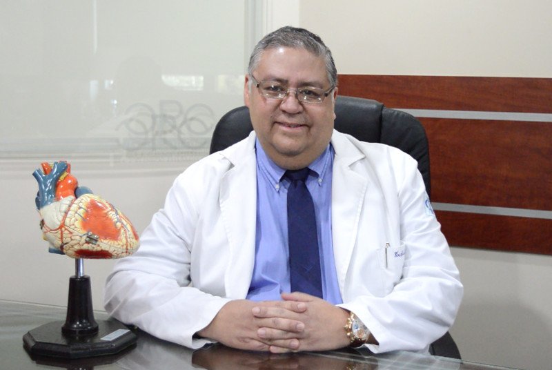
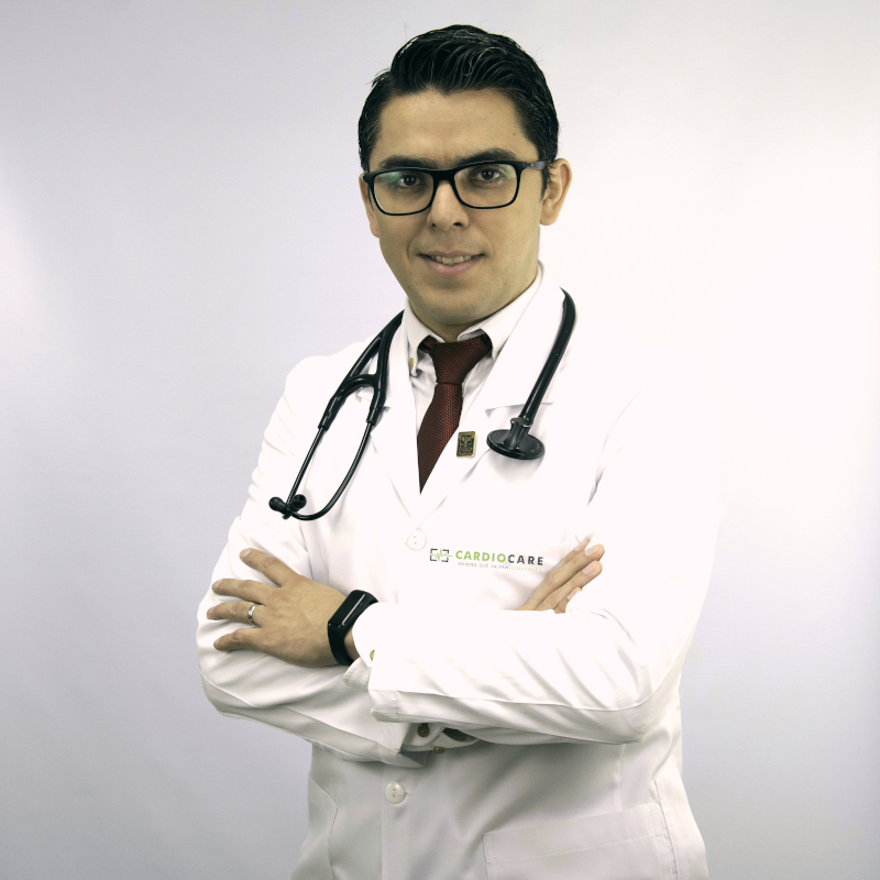
Dr. Ricardo Muñoz
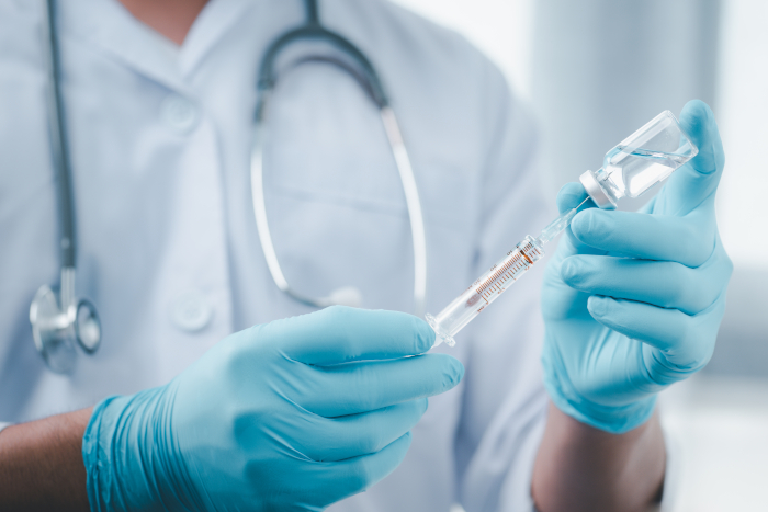
Consulta médica cardiológica | Hemodinamia |Ecocardiografia adultos | Prueba de esfuerzo | Monitor...
Ver Más...
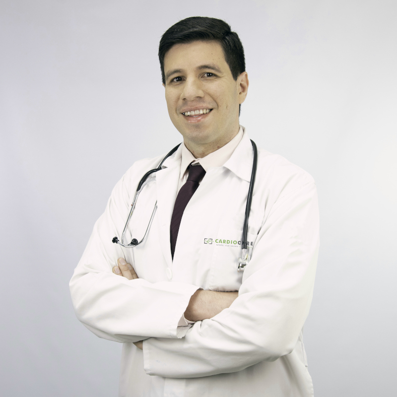
Dr. Jose Luis Alvarado
Consulta médica cardiológica | Tilt test | Prueba de esfuerzo | Cardiólogos Guatemala | Médicos ...
Ver Más...
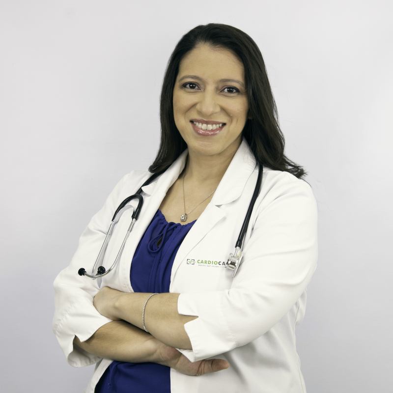
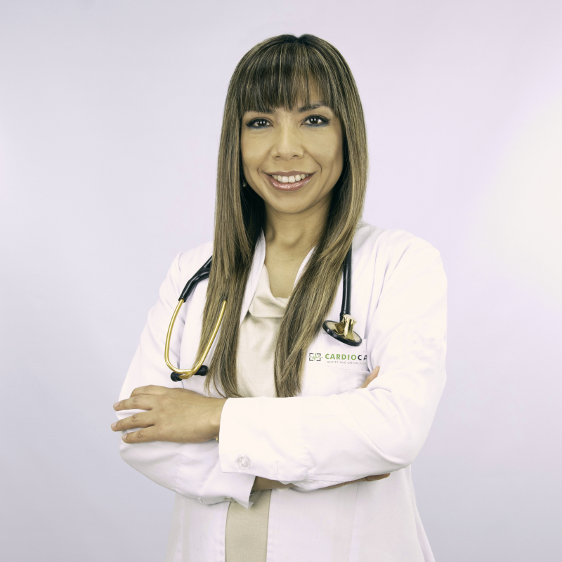
Dra. Beatriz Domínguez
Consulta médica cardiología | Ecocardiograma adultos | Eco Stress con dobutamina | Stress Eco en B...
Ver Más...
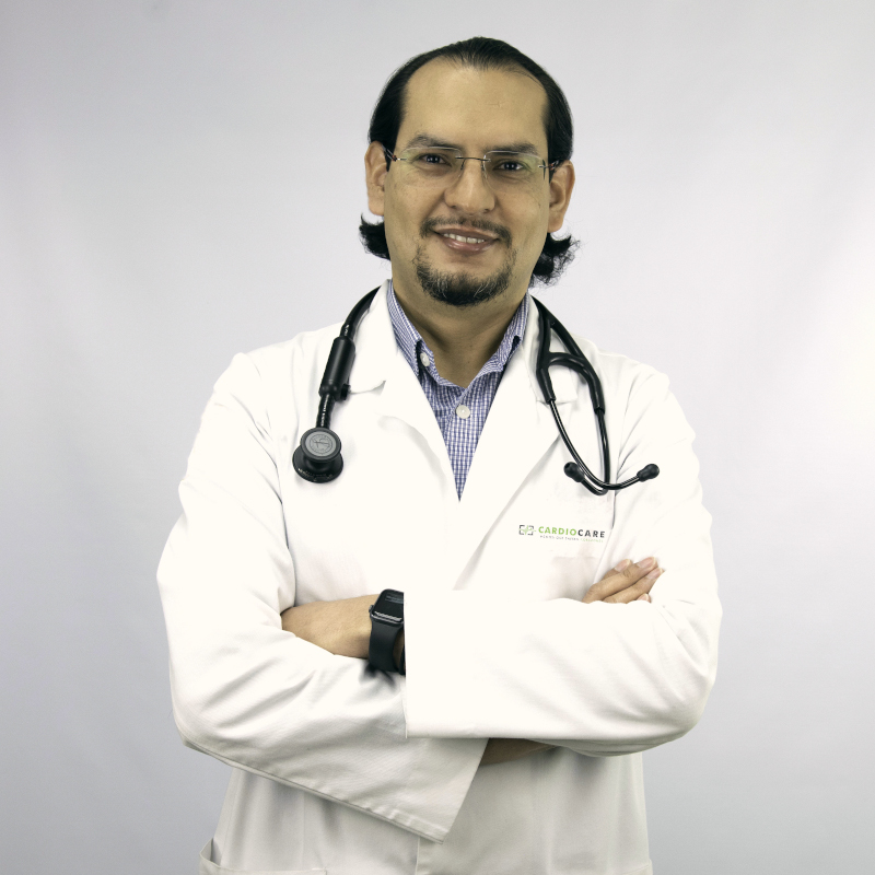
Dr. Steven Gil
Consulta médica cardiológica | Ecocardiografía adultos | Stress Eco con Dobutamina | Prueba de es...
Ver Más...
Dr. Julio Guillén
Consulta médica cardiológica | Clínica de marcapasos | Arritmias | Hemodinamia | Prueba de esfuer...
Ver Más...
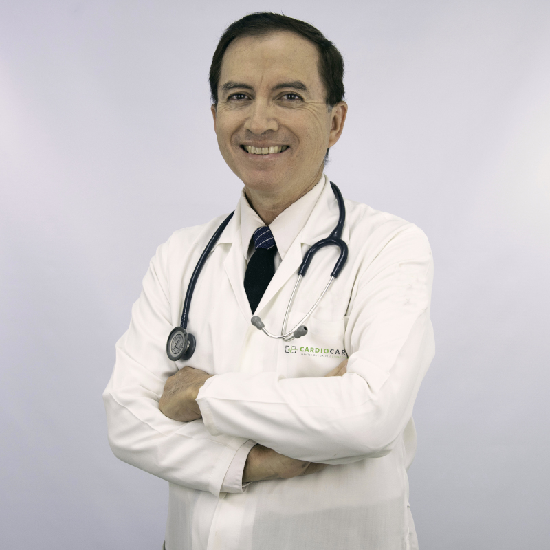
Dr. César López
Consulta médica cardiológica | Ecocardiografía adultos | Prueba de esfuerzo | Rehabilitación car...
Ver Más...
Dr. Walter Mancilla
Consulta cardiología pediátrica | Ecocardiografía pediátrica | Monitoreo holter de 24 horas pedi...
Ver Más...
Dr. Héctor Mora
Consulta médica cardiológica | Prueba de esfuerzo | MAPA | Holter | Cardiólogos Guatemala | Médi...
Ver Más...
Dra. Norma Ordoñez
Consulta médica cardiológica | Ecocardiografía adultos | Strain Eco | Prueba de esfuerzo | Tilt t...
Ver Más...
Dr. José Carlos Penagos
Ecocardiograma adultos | Eco Stress con dobutamina | Stress Eco en Banda |Tilt test | Consulta médi...
Ver Más...
Dr. Ery Mario Rodríguez
Consulta médica de flebología | Doppler venoso y arterial de miembros |Safenectomía |Flebectomí...
Ver Más...
Dr. Héctor Santos Carranza
Consulta médica cardiológica | Tilt test | Prueba de esfuerzo | Cardiólogos Guatemala | Médicos ...
Ver Más...
Dr. Harry Soto
Consulta médica cardiológica |Ecocardiografía adultos | Prueba de esfuerzo |Tilt test | Cardiólo...
Ver Más...
Dr. Luis Velásquez
Ecocardiograma adultos | Eco Stress con dobutamina | Stress Eco en Banda |Doppler carotídeo | Eco t...
Ver Más...
Dr. Salvador Aguilar
Consulta médica cardiológica |Ecocardiografia adultos | Prueba de esfuerzo | Monitoreo de presión...
Ver Más...
Dr. Lester Rojas
Consulta médica cardiológica |Prueba de esfuerzo | Monitoreo de presión arterial | Monitoreo holt...

Dr. Gerardo Arévalo
Consulta médica cardiológica | Clínica de marcapasos | Arritmias | Hemodinamia | Prueba de esfuer...
Ver Más...

Dr. Pablo Pérez
Consulta médica cardiológica |Ecocardiografia adultos | Prueba de esfuerzo | Monitoreo de presión...
Ver Más...

Dr. Romeo Guevara
Cirugías |Cirugía vascular | Cirugía endovascular| Angiólogo | Cardiólogo Guatemala | Médicos ...
Ver Más...

Dr. Benjamín Godínez
Consulta médica cardiológica | Tilt test | Prueba de esfuerzo | Cardiólogos Guatemala | Médicos ...
Ver Más...

Dra. Josselin Morales
Consulta médica nefrología | Nefrólogos Guatemala | Médicos del corazón
Ver Más...